What is tissue viability?
According to the Tissue Viability Society 2009):
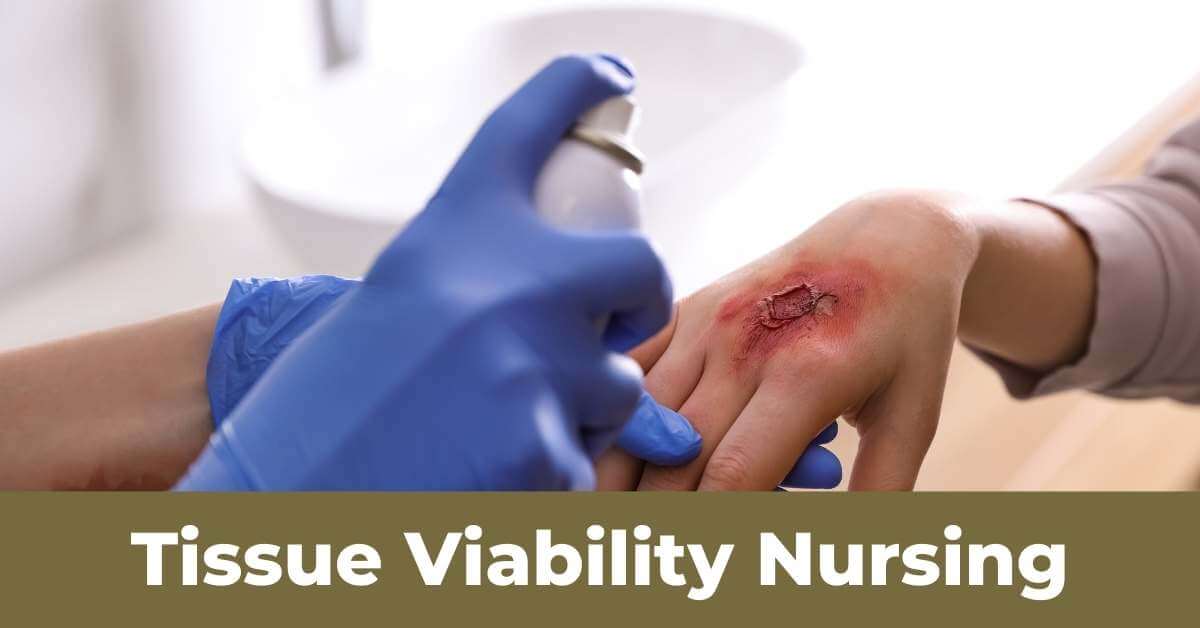
Tissue viability is a developing field that primarily focuses on all facets of skin and soft tissue wounds, such as acute surgical wounds, pressure ulcers, and all types of leg ulceration.
The Tissue Viability Nurses provide guidance and assistance in a variety of ways to staff members, and adult and child patients. This involves assisting inpatients and patients cared for by district nurses and practice nurses in the larger community, as well as assisting and advising care facilities.
What is the purpose of tissue viability?
Tissue Viability helps patients who have chronically unhealed wounds like bed sores, leg ulcers, diabetic wounds, and other wounds.
The Tissue Viability nurse assesses and treats patients with difficult non-healing wounds, such as leg ulcers, pressure ulcers, fungating wounds, and post-operative wounds.
Guidelines on tissue viability and wound management
Health Care Professionals (HCPs) treat a wide range of wounds that might be brought on by trauma, surgical procedures, or disease processes. When complex interventions are essential, there is also a variety of treatment modalities available to the HCP. However, in most cases, these heal naturally without the need for them.
Regardless of the employer, guidelines on tissue viability and wound management are essential for healthcare professionals providing wound care since they outline the minimal level of care that should be provided. Following this advice will guarantee that all wound care is provided is based on recognised, evidence-based care standards to get the best results for patients.
The Physiological Process of Wound Healing
There are two main types of healing, primary intention, and secondary intention.
Primary intention
These are clean, straightforward wounds with little tissue loss and edges that can be stitched, clipped, glued, etc. to hold them together. With epithelial continuity, they recover rather fast and within 48 hours.
Secondary intention
These are more complicated and have excessive tissue loss. The edges cannot be brought together. The wounds are ‘open’ and take much longer to heal. All tissues in the body are capable of healing through regeneration (replication of cells) and repair (connective tissue replaces damaged tissue).
Healing by Secondary Intention
There are four distinct stages of wound healing. These phases will overlap, and the length of time it takes to move through them will vary and rely on a variety of factors, including age and overall health. Most of the time, until the wound is epithelialized, there are no distinct transitions or gaps between stages; instead, both inflammatory and proliferative processes coexist at variable intensities.
The Vascular Response
Within seconds of injury, the blood vessels injured end contract to reduce blood flow and start the clotting process. This is accelerated by platelet aggregation and the release of numerous growth factors required for wound healing. Fibrin mesh blood clot forms, trapping the blood cells and sealing the wound. White blood cells and inflammatory stage chemicals are drawn to the site by the growth factors generated during vasodilatation of the arteries surrounding the lesion.
The Inflammatory Phase
Localized indications of erythema, heat, and oedema are present due to increased blood flow to the region and fluid buildup in the soft tissue. The sensory nerve is subjected to pressure, which results in discomfort and movement restrictions. Within hours of an injury, neutrophils reach the wound site and offer the first anti-infection defense.
These are phagocytic and engulf foreign bodies, having a short life being replaced by Monocytes (also phagocytic) but these develop into macrophages and play an important role in the wound healing process. Clean wounds will spend up to 36 hours in the inflammatory phase, but the process is prolonged if the wound becomes necrotic or infected.
The inflammatory response may be suppressed or absent in patients who are immunosuppressed, for example, people with HIV or AIDS and those receiving immunosuppressive drugs such as NSAIDS, cytotoxics, or steroids. Therefore, they may fail to activate the normal healing process.
The Proliferation Phase
At this point, connective tissue starts to fill the incision. Granulation, which promotes the growth of new connective tissue, is the term used to describe the generation of new capillary growth in the wound bed. The granular and slightly uneven appearance of granulation tissue helps to identify it. Contraction also takes place as connective tissue is being created. When myofibroblasts gather around the border of the incision, they might contract, drawing the edges of the wound together and shrinking the size of the wound.
The Maturation Phase
The wound has now healed due to the growth of connective tissue and epithelization. When macrophages stimulate a scar, it changes until a stronger scar form as the collagen produced beneath the skin is organized into a more robust structure. Compression can lessen the build-up of scar formation and ultimately result in a flatter scar as the scar ages and the blood supply declines. The scar will, at most, only be 80% as robust as healthy tissue.
The bottom line
Tissue viability is an essential part of wound management. Tissue viability Nurses deliver advice and support to both adult and child patients, as well as staff, in several different ways. This includes supporting inpatients and patients cared for in the wider community by district nurses, and practice nurses, and providing help and advice to care homes.
